Center for Macromolecular Interactions

The Center for Macromolecular Interactions (CMI) in the department of Biological Chemistry and Molecular Pharmacology at Harvard Medical School has a mission to enhance basic research in the HMS community by providing scientific consultation, training and access to shared biophysical instruments for the characterization and analysis of macromolecules and their complexes. 

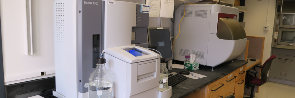
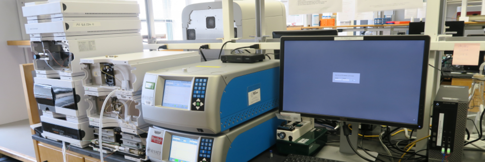
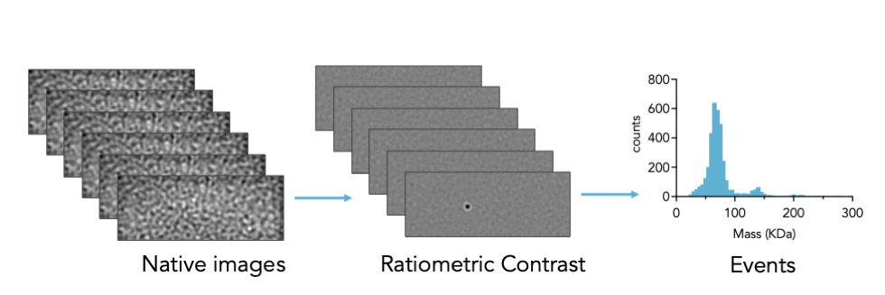
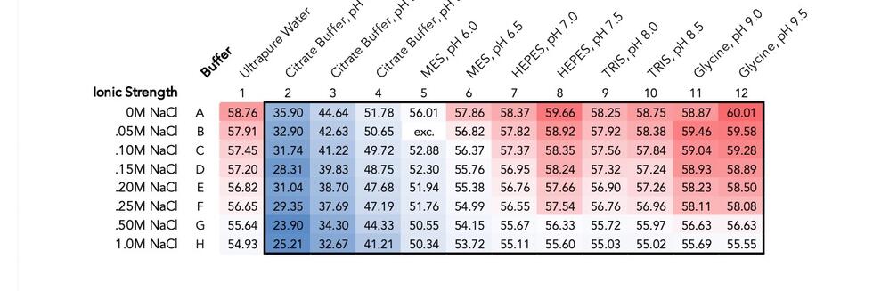
The facility currently offers training and access to instruments for Isothermal Titration Calorimetry (ITC)Surface Plasmon Resonance (SPR)Biolayer Interferometry (BLI)MicroScale Thermophoresis (MST)Differential Scanning Fluorimetry (DSF)Circular Dichroism (CD), and Light Scattering: including size-exclusion chromatography with multi-angle light scattering (SEC-MALS), Dynamic Light Scattering (DLS), and Mass Photometry (MP)

The CMI offers limited data collection services for the characterization of protein secondary structure, mass and oligomeric state, polydispersity, aggregation state, hydrodynamic radius, and thermal stability.  Using a library developed in the lab of Andrew Kruse, the CMI is also offering yeast surface display nanobody selections services.

Acknowledgements

Researchers using CMI equipment or services agree to acknowledge the support of "the Center for Macromolecular Interactions at Harvard Medical School" in publications arising from use of the facility. If a CMI staff member has made an important intellectual contribution to the project, as determined by generally accepted criteria for academic collaborations, then that person should be considered for authorship on the publication. 

CMI users may enter publications directly in the CMI PPMS system or email the Pubmed ID (or the reference) to cmi@hms.harvard.edu.  

Recent CMI User Publications

Sharma P, Zhang X, Ly K, Zhang Y, Hu Y, Ye AY, Hu J, Kim JH, Lou M, Wang C, Celuzza Q, Kondo Y, Furukawa K, Bundle DR, Furukawa K, Alt FW, Winau F. The lipid globotriaosylceramide promotes germinal center B cell responses and antiviral immunity. Science 2024;383(6684):eadg0564.Abstract
Influenza viruses escape immunity owing to rapid antigenic evolution, which requires vaccination strategies that allow for broadly protective antibody responses. We found that the lipid globotriaosylceramide (Gb3) expressed on germinal center (GC) B cells is essential for the production of high-affinity antibodies. Mechanistically, Gb3 bound and disengaged CD19 from its chaperone CD81, permitting CD19 to translocate to the B cell receptor complex to trigger signaling. Moreover, Gb3 regulated major histocompatibility complex class II expression to increase diversity of T follicular helper and GC B cells reactive with subdominant epitopes. In influenza infection, elevating Gb3, either endogenously or exogenously, promoted broadly reactive antibody responses and cross-protection. These data demonstrate that Gb3 determines the affinity and breadth of B cell immunity and has potential as a vaccine adjuvant.
Stinson BM, Carney SM, Walter JC, Loparo JJ. Structural role for DNA Ligase IV in promoting the fidelity of non-homologous end joining. Nat Commun 2024;15(1):1250.Abstract
Nonhomologous end joining (NHEJ), the primary pathway of vertebrate DNA double-strand-break (DSB) repair, directly re-ligates broken DNA ends. Damaged DSB ends that cannot be immediately re-ligated are modified by NHEJ processing enzymes, including error-prone polymerases and nucleases, to enable ligation. However, DSB ends that are initially compatible for re-ligation are typically joined without end processing. As both ligation and end processing occur in the short-range (SR) synaptic complex that closely aligns DNA ends, it remains unclear how ligation of compatible ends is prioritized over end processing. In this study, we identify structural interactions of the NHEJ-specific DNA Ligase IV (Lig4) within the SR complex that prioritize ligation and promote NHEJ fidelity. Mutational analysis demonstrates that Lig4 must bind DNA ends to form the SR complex. Furthermore, single-molecule experiments show that a single Lig4 binds both DNA ends at the instant of SR synapsis. Thus, Lig4 is poised to ligate compatible ends upon initial formation of the SR complex before error-prone processing. Our results provide a molecular basis for the fidelity of NHEJ.
Hauser BM, Sangesland M, Lam EC, St Denis KJ, Sheehan ML, Vu ML, Cheng AH, Sordilla S, Lamson DT, Almawi AW, Balazs AB, Lingwood D, Schmidt AG. Heterologous Sarbecovirus Receptor Binding Domains as Scaffolds for SARS-CoV-2 Receptor Binding Motif Presentation. ACS Infect Dis 2024;10(2):553-561.Abstract
Structure-guided rational immunogen design can generate optimized immunogens that elicit a desired humoral response. Design strategies often center on targeting conserved sites on viral glycoproteins that will ultimately confer potent neutralization. For SARS-CoV-2 (SARS-2), the surface-exposed spike glycoprotein includes a broadly conserved portion, the receptor binding motif (RBM), that is required to engage the host cellular receptor, ACE2. Expanding humoral responses to this site may result in a more potent neutralizing antibody response against diverse sarbecoviruses. Here, we used a "resurfacing" approach and iterative design cycles to graft the SARS-2 RBM onto heterologous sarbecovirus scaffolds. The scaffolds were selected to vary the antigenic distance relative to SARS-2 to potentially focus responses to RBM. Multimerized versions of these immunogens elicited broad neutralization against sarbecoviruses in the context of preexisting SARS-2 immunity. These validated engineering approaches can help inform future immunogen design efforts for sarbecoviruses and are generally applicable to other viruses.
Chanez-Paredes SD, Abtahi S, Zha J, Li E, Marsischky G, Zuo L, Grey MJ, He W, Turner JR. Mechanisms underlying distinct subcellular localization and regulation of epithelial long myosin light-chain kinase splice variants. J Biol Chem 2024;300(2):105643.Abstract
Intestinal epithelia express two long myosin light-chain kinase (MLCK) splice variants, MLCK1 and MLCK2, which differ by the absence of a complete immunoglobulin (Ig)-like domain 3 within MLCK2. MLCK1 is preferentially associated with the perijunctional actomyosin ring at steady state, and this localization is enhanced by inflammatory stimuli including tumor necrosis factor (TNF). Here, we sought to identify MLCK1 domains that direct perijunctional MLCK1 localization and their relevance to disease. Ileal biopsies from Crohn's disease patients demonstrated preferential increases in MLCK1 expression and perijunctional localization relative to healthy controls. In contrast to MLCK1, MLCK2 expressed in intestinal epithelia is predominantly associated with basal stress fibers, and the two isoforms have distinct effects on epithelial migration and barrier regulation. MLCK1(Ig1-4) and MLCK1(Ig1-3), but not MLCK2(Ig1-4) or MLCK1(Ig3), directly bind to F-actin in vitro and direct perijunctional recruitment in intestinal epithelial cells. Further study showed that Ig1 is unnecessary, but that, like Ig3, the unstructured linker between Ig1 and Ig2 (Ig1/2us) is essential for recruitment. Despite being unable to bind F-actin or direct recruitment independently, Ig3 does have dominant negative functions that allow it to displace perijunctional MLCK1, increase steady-state barrier function, prevent TNF-induced MLCK1 recruitment, and attenuate TNF-induced barrier loss. These data define the minimal domain required for MLCK1 localization and provide mechanistic insight into the MLCK1 recruitment process. Overall, the results create a foundation for development of molecularly targeted therapies that target key domains to prevent MLCK1 recruitment, restore barrier function, and limit inflammatory bowel disease progression.
Lim S, Reilly CB, Barghouti Z, Marelli B, Way JC, Silver PA. Tardigrade secretory proteins protect biological structures from desiccation [Internet]. bioRxiv 2023; Publisher's VersionAbstract
Tardigrades, microscopic animals that survive a broad range of environmental stresses, express a unique set of proteins termed tardigrade-specific intrinsically disordered proteins (TDPs). TDPs are often expressed at high levels in tardigrades upon desiccation, and appear to mediate stress adaptation. Here, we focused on the proteins belonging to the secretory family of tardigrade proteins termed secreted-abundant heat soluble (“SAHS”) proteins, and investigated their ability to protect diverse biological structures. Recombinantly expressed SAHS proteins prevented desiccated liposomes from fusion, and enhanced desiccation tolerance of E. coli and Rhizobium tropici upon extracellular application. Molecular dynamics simulation and comparative structural analysis suggest a model by which SAHS proteins may undergo a structural transition upon desiccation, in which removal of water and solutes from a large internal cavity in SAHS proteins destabilizes the beta-sheet structure. These results highlight the potential application of SAHS proteins as stabilizing molecules for preservation of cells.Competing Interest StatementThe authors have declared no competing interest.
More

CMI News

CMI FAQs

February 26, 2024

The CMI Knowledge Base is a place to find answers to frequently asked questions about the CMI and CMI resources.

If you have a question or a suggestion for our FAQ, contact us.

New Jasco J-1500 instrument installed

April 5, 2023

We are excited to announce that the Jasco J-1500 has been installed. The J-815 is being retired.  Active CD user rights have been transferred to the new instrument, as new training will not be required.

A new CMI User guide for the Jasco J-1500 is available.  The SpectraManager2 software and most of the parameter settings are the same.  Let us know if you need help getting started.

More