Multimode Plate Reader
CMI Spark resources and user guide⬇︎
Multimode plate readers are used for measuring fluorescence, absorbance or luminescence in a microplate and support a wide array of assays. The CMI has a Spark Multimode Plate Reader from Tecan, configured with Absorbance mode, Luminescence mode, and enhanced Fusion Optics Fluorescence mode.

Fusion Optics supports flexible set-up of fluorescence experiments using monochromator-based optics, filter-based optics, or a combination. Filters allow for higher sensitivity and speed and monochromators allow for greater flexibility and specificity in wavelength selection.
Key Features and Applications
- Fluorescence (enhanced)
- Top and bottom read – End point and spectra
- Fusion Optics
- Monochromator spectral range: Ex. 230-900 nm; Em. 280 – 900 nm
- Excitation and Emission Filters: 12 common filters (see specifications)
- Fluorescence Polarization (FP)
- Fluorescence Resonance Energy Transfer (FRET)
- Time-resolved fluorescence (TRF)
- Absorbance
- Top read – Endpoint and UV/Vis spectra
- spectral range: 200 – 1000 nm
- Luminescence (enhanced)
- Top read – glow, flash, multi-color, spectra
- spectral range: 370 – 700 nm
- NOT configured for AlphaScreen, Fluorescence Imaging or Cell-based assays.
Fluorescence Polarization (FP)
Fluorescence Polarization (FP), also called Fluorescence Anisotropy, measures the rotational mobility of fluorescently labeled molecules in solution. When a sample is excited with plane-polarized light, only those fluorophores aligned with the light’s electric vector become excited. If the labeled molecule rotates slowly (typically because it's bound to a larger partner), the emitted light retains much of the original polarization. If the molecule is small or unbound and rotates rapidly, the polarization decreases as the emission becomes depolarized.
FP is widely used to monitor binding interactions, such as protein–protein, protein–DNA, or small molecule binding assays. High polarization values indicate binding (reduced rotational mobility), while low values indicate free, unbound molecules. The method is homogeneous (no separation/washing required), rapid, and suitable for high-throughput screening.

SparkControl will measure both parallel and perpendicular fluorescence intensities for each well.
Polarization (mP or P):
\[P=\frac{I_{\mid\mid}-I_\bot}{I_{\mid\mid}+I_\bot}\]Anisotropy (r):
\[r=\frac{I_{\mid\mid}-I_\bot}{I_{\mid\mid}+2I_\bot}\]
Where I|| is intensity of parallel and I⟂ is perpendicular emission.
| Fluorophore | τf (ns) | Optimal Mass*(kDa) | Usable Mass Range (kDa) | Ex (nm) | Em (nm) |
|---|---|---|---|---|---|
Cy3 | 0.5 | 0.35 | <1–10 | 550 | 570 |
Cy5 | 1 | 0.98 | <1–15 | 650 | 670 |
Tetramethyl- rhodamine | 2.5 | 3 | <10–50 | 555 | 580 |
Fluorescein FITC | 4 | 6 | <10–75 | 495 | 520 |
AlexaFluor 488 | 4.1 | 6.5 | <10–75 | 495 | 519 |
BODIPY TMR | 5.4 | 9.8 | <10–100 | 544 | 574 |
BODIPY FL | 5.5 | 10 | <10–100 | 503 | 512 |
Dansyl | ~20 | 54 | <50-300 | 340 | 520 |
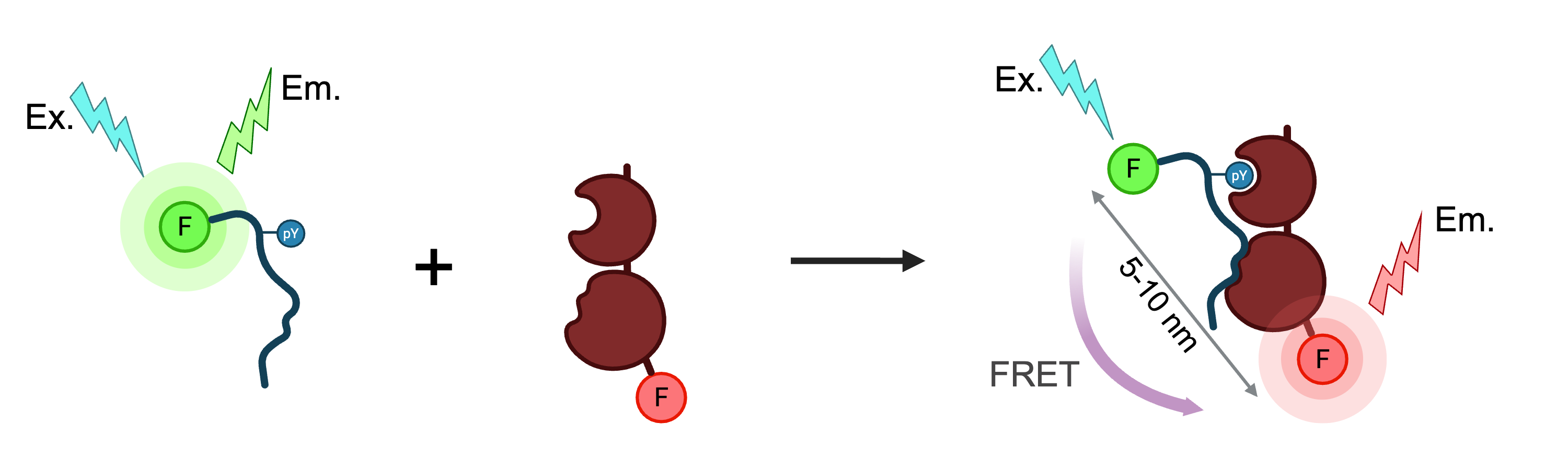
Förster Resonance Energy Transfer (FRET)
Förster Resonance Energy Transfer (FRET) is a mechanism describing the transfer of energy between two fluorophores. The donor molecule, absorbs light energy and, if the acceptor molecule is close enough, can transfer this energy without the emission of light (non-radiatively). This energy transfer, which occurs through a dipole-dipole coupling, then causes the acceptor to emit its own fluorescence.
The efficiency of this energy transfer is very sensitive to the distance between the donor and acceptor, decreasing proportionally to the sixth power of the distance separating them. This makes FRET an extremely precise "spectroscopic ruler" for measuring nm distances. For FRET to occur, several conditions must be met:
- Proximity: The donor and acceptor must be very close, typically within 1-10 nanometers of each other.
- Spectral Overlap: The emission spectrum of the donor fluorophore must overlap with the absorption spectrum of the acceptor.
- Dipole Orientation: The orientation of the donor's emission dipole and the acceptor's absorption dipole must be roughly parallel.
A key parameter in FRET is the Förster radius (R₀), which is the distance at which the energy transfer efficiency is 50%. This value helps determine the feasible range of distances that can be measured with a specific donor-acceptor pair.
Applications of FRET:
- Detecting and tracking interactions between proteins.
- Measuring distances between different parts of a single protein to understand its conformation and folding.
- Studying DNA and RNA structures.
Limitations of FRET:
- Standard FRET is its highly susceptibility to background noise from scattered excitation light and natural fluorescence from other molecules in the sample (autofluorescence).
| Donor | Ex (nm) | Em (nm) | Acceptor | Ex (nm) | Em (nm) |
|---|---|---|---|---|---|
CFP (Cyan FP) | 433 | 475 | YFP (Yellow FP) | 514 | 527 |
GFP (Green FP) | 488 | 509 | YFP (Yellow FP) | 514 | 527 |
FITC (Fluorescein) | 495 | 519 | TRITC (Tetramethylrhodamine) | 557 | 576 |
Cy3 | 550 | 570 | Cy5 | 649 | 670 |
Alexa Fluor 488 | 495 | 519 | Alexa Fluor 555 | 555 | 565 |
GFP (Green FP) | 488 | 509 | mCherry | 587 | 610 |
Cy3 | 550 | 570 | Cy3.5 | 581 | 596 |
FAM (Carboxyfluorescein) | 495 | 520 | Texas Red | 595 | 615 |
Cy3.5 | 581 | 596 | Cy5.5 | 675 | 694 |
Alexa Fluor 546 | 556 | 573 | Alexa Fluor 647 | 650 | 668 |
B-Phycoerythrin | 545 | 575 | Cy5 | 649 | 670 |
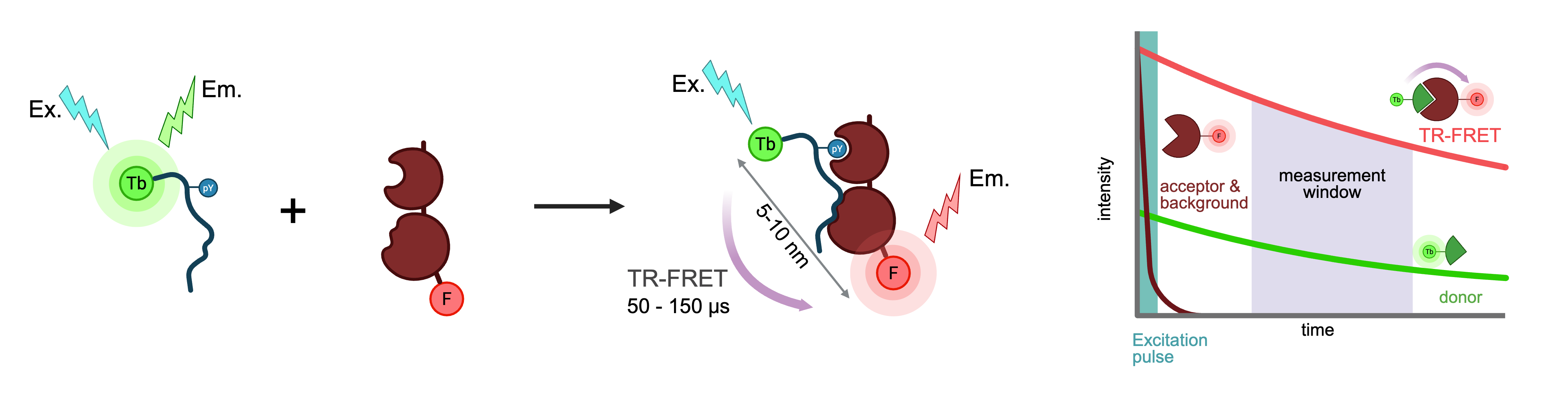
Time-Resolved Förster Energy Transfer (TR-FRET), also called Time-Resolved Fluorescence Resonance Energy Transfer, is a highly sensitive and robust biochemical technique used to study molecular interactions. It combines the principles of FRET (measuring energy transfer between two light-sensitive molecules, or fluorophores) with Time-Resolved Fluorescence (measuring fluorescence signals after a specific delay). This unique combination dramatically reduces background noise and interference, making it a standard for high-throughput screening (HTS) in drug discovery and for quantifying biomolecular interactions. FRET occurs when a donor (excited by a light source) transfers energy non-radiatively to a nearby acceptor, but only if they are within a 1–10 nm proximity. This results in quenching of donor fluorescence and emission of light from the acceptor. Time-Resolved-FRET, uses a long-lifetime fluorophore as the donor. After excitation, a delay is introduced (~50–150 μs) before the emission signal is detected. This delay allows short-lived background fluorescence from the sample matrix or autofluorescence to decay, resulting in a much lower background and higher assay sensitivity.
- Donor Fluorophore - a lanthanide chelate (Europium or Terbium). Lanthanides have an very long fluorescence lifetime (milliseconds), whereas typical background fluorescence lasts only for nanoseconds.
- Acceptor Fluorophore - a conventional fluorophore (e.g., Alexa Fluor, Cy5, or specialized proprietary dyes) that can accept energy from the donor.
Advantages
- Low Background Signal. Due to time-resolved measurement, native fluorescence and other short-lived signals are excluded.
- Sensitivity. Capable of detecting low-affinity or transient interactions.
- Versatility. Suitable for a wide range of applications including protein-protein, protein-peptide, and small molecule binding studies.
| Fluorophore | CoraFluor 1 | LanthaScreen | LANCE |
|---|---|---|---|
Supplier | Tocris Bioscience | Thermo Fisher Scientific | Revvity |
Donor Chemistry | Terbium chelate | Terbium (Tb³⁺) chelate | Europium (Eu³⁺) chelate/cryptate |
Excitation Wavelength | 337 nm | 340 nm (typical) | 320–340 nm |
Emission Bands | 490, 545, 590, 620 nm (Tb³⁺) | 495, 545, 570, 620 nm (Tb³⁺) | 615 nm (Eu³⁺); long-lived |
Compatible Acceptors | FAM (fluorescein), TMR, Cy5, GFP, mCherry, Alexa Fluor™ 488 | Alexa Fluor 488, 546, 594, 647, FAM, GFP, Cy5, others | ULight (Revvity proprietary), Cy5, APC, Alexa Fluor™ 647 |
Reactive Chemistries | Amine-reactive (PFP ester), Thiol-reactive (maleimide), Haloalkane | Amine-reactive (NHS ester), protein labeling kits | Amine-reactive (ITC chelate), protein labeling kits |
Key Features | High brightness/stability Multiple reactive forms Best for in vitro/cell-free assays | Widely compatible, robust kits multiple acceptor choices optimized for screening | Sensitive, robust long-lived fluorescence low background |
Product Page |
Spark Plate Reader Resources
CMI Tecan Spark Multimode Plate Reader User Guide - NEW
Please let us know if you find errors.
Spark Multimode Plate Reader from Tecan.
Instrument Specifications
Tecan Spark
Temperature Control: Ambient +3C – 42C
Plate Formats: 96-well, 384-well, 1536-well